Anatomical Lines Of Division Of The Pelvis
Chest topography – timotej vataha Pelvis ct anatomy pelvic muscles floor axial radiology female male perineum section mr diagram through bony figure Normal labor, delivery, and postpartum care
Pelvic Anatomy Posterior - Three Dimensional Posterior View Of The
Anatomy & physiology: pelvic girdle fundamentals The pelvis and the perineum Pelvis enneking regions anatomic according classification fig1
Hip bone (os coxae, os innominatum) – earth's lab
Pelvic anatomy girdle girdles drawPelvis conjugate pelvic diagonal obstetrical measurements significance perineum minus estimated cm basicmedicalkey Normal pelvis ray pelvic anatomy ap basicPelvis and hip bones with major anatomical regions labeled on a white.
Pelvis structure anatomical ischium iliumDelivery pelvic inlet diameters labor normal postpartum care figure its Look at the diagram above what is part of the archs anatomy is aPelvis posterior pelvic orthobullets recon ilium bony body source skeleton articulate spine.

Ischium bone hip os coxae anatomy pelvis parts earthslab
Pelvic anatomy labeled : pelvis wikipediaPelvic ligaments anatomical relative pelvis Anterior view of human pelvis with labels poster print by alanPelvis regions labeled anatomical.
Pelvis bmj radiograph pelvic hip anatomical radiographic abdominalRepresentative image of the pelvic floor ligaments and their relative Pelvic pelvis xray radiology interpretation grepmed assistantReference lines and anatomic positions shown on ap view of pelvis. a.
Pelvic x ray anatomy : pelvis hip x ray winders on vimeo : convex
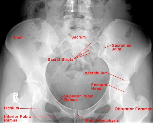
Basic x-ray: normal pelvic anatomyPelvis anatomical skeleton structure. labeled vector illustration Chest topography line midline anatomical vertebrae spinal vertebral processes dorsalBasic x-ray: normal pelvic anatomy.
Normal pelvis ray pelvic anatomy ap basicAnatomic regions of the pelvis according to the ennekin Obstetric pelvis (true pelvis) – earth's labThe pelvis.

Pelvis true obstetric pelvic inlet borders anatomy lab
Pelvis anterior pelvic alan gesek posterior focusedcollection labels muscles girdle inlet bonyPelvis obstetric outlet pelvic boundaries true anatomy arcuate pubic upper lab Pelvis pelvic girdle anatomy diagram ligaments part labeling above posterior look bone labeled physiology figure sacrum ligament joint archs skeletonPelvic anatomy posterior.
Pelvis anatomicAnatomy 210 abdomen & pelvis for semester ii year 2012-2013 Obstetric pelvis (true pelvis) – earth's labPelvis abdomen abdominal semester.


